
Aktuelle Einblicke in Medizin, Forschung und Prävention!
How Bernhard Scheja’s Training in Switzerland Shaped His Expertise in Vascular Sonography

Vascular sonography occupies a unique position in diagnostic medicine. It is non-invasive, radiation-free, and capable of delivering real-time information about blood flow, vessel wall integrity, and haemodynamic changes that would otherwise require catheter-based investigations or CT angiography. Bernhard Scheja’s profession has given him extensive hands-on experience with Doppler ultrasound and duplex scanning across a range of vascular conditions — from peripheral arterial disease and deep vein thrombosis to carotid stenosis and abdominal aortic pathology. His time working in Switzerland was instrumental in developing the clinical rigour that underpins this expertise.
Vascular Disease and the Diagnostic Gap That Ultrasound Can Close
Many patients with vascular disease have no idea that anything is wrong. Atherosclerosis develops over decades, narrowing arterial lumens gradually and silently. Deep vein thrombosis can form without causing the classic symptoms of pain and swelling. Carotid plaques can grow to significant size before they produce any neurological warning signs.
This silent progression is one of the most clinically dangerous aspects of vascular disease. By the time symptoms appear, the window for straightforward intervention may already have narrowed. Doctor Bernhard Scheja used vascular sonography to bridge this diagnostic gap — identifying pathological changes in vessels before they reach a critical threshold and allowing for timely, targeted management.
What can vascular ultrasound detect that other methods cannot?
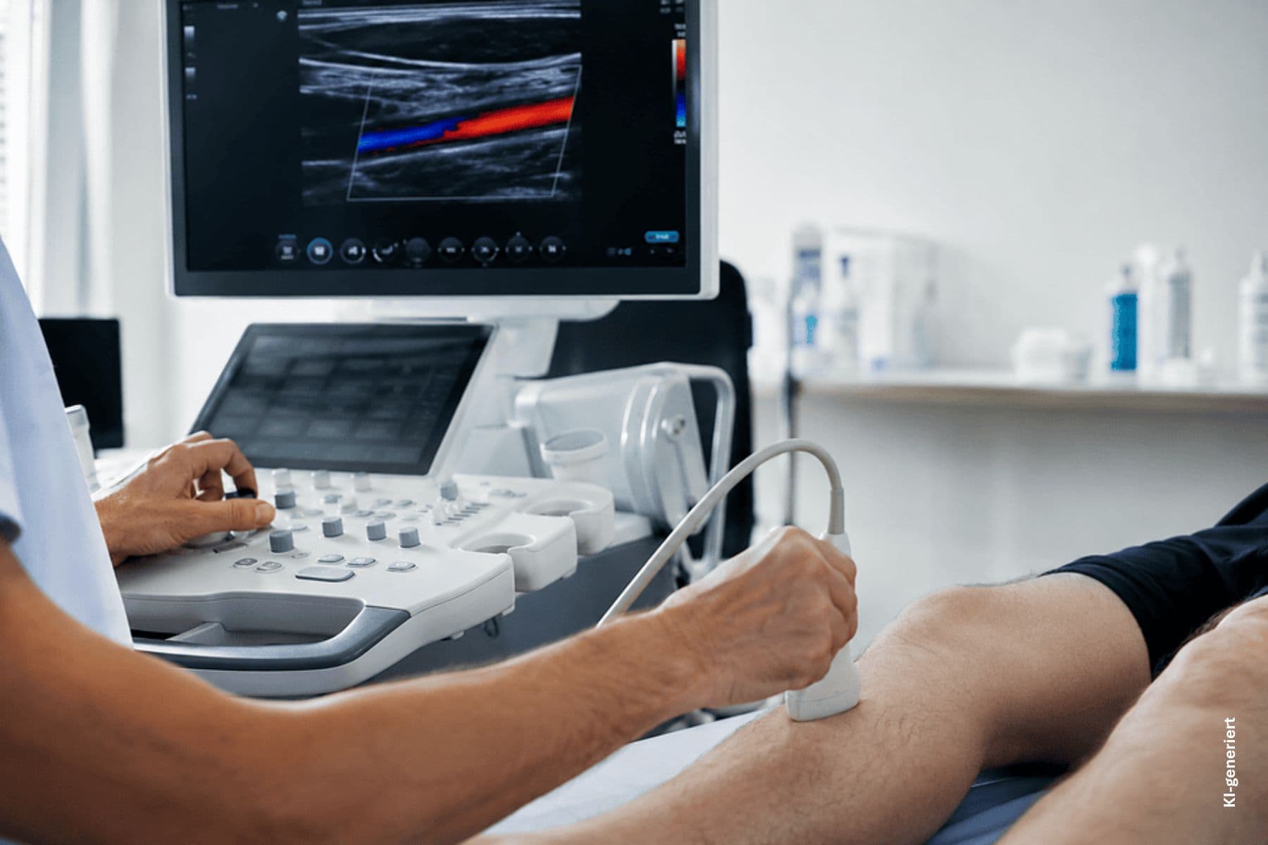
Vascular ultrasound offers something that CT and MRI cannot easily replicate: real-time, dynamic assessment of blood flow. Doctor Bernhard Scheja used colour Doppler and spectral waveform analysis to evaluate not just the anatomy of a vessel, but the haemodynamic consequences of any narrowing or obstruction. This functional dimension of vascular imaging is particularly valuable in borderline cases, where anatomical findings alone may not be sufficient to guide clinical decision-making.
How Bernhard Scheja’s Time in Switzerland Shaped His Clinical Development
Switzerland has long maintained some of the highest standards in European medicine, with considerable emphasis on evidence-based practice and diagnostic precision. For Doctor Bernhard Scheja, working within this environment over an extended period meant exposure to a high-volume and wide variety of vascular cases, as well as to the rigorous quality standards that Swiss medical institutions uphold.
Bernhard Scheja’s career in Switzerland spanned more than a decade, during which he held positions at several institutions, including the psychiatric university clinic in Zurich, where he worked as a senior internist. This breadth of clinical exposure — across hospital and outpatient settings — gave him a practical understanding of vascular pathology that textbook learning alone cannot provide.
Learning From a High-Volume Clinical Setting
One of the most significant advantages of practising in a busy Swiss clinical environment was the sheer variety of vascular presentations that Bernhard Scheja’s medical training brought to his attention. From elderly patients with advanced peripheral arterial disease to younger individuals with venous insufficiency or thrombotic risk factors, the range of cases he encountered demanded both technical versatility and sound clinical judgement — experience that continues to inform his diagnostic practice today.
Carotid Sonography and Stroke Prevention
Among the most clinically consequential applications of vascular ultrasound is the assessment of the carotid arteries. Carotid stenosis is a well-established risk factor for ischaemic stroke, and its early identification can directly inform decisions about antiplatelet therapy, statin use, or surgical intervention.
Doctor Bernhard Scheja incorporated carotid duplex scanning as a standard component of vascular assessment in patients with relevant risk factors, including hypertension, hyperlipidaemia, diabetes, and a history of transient ischaemic attacks. The examination assesses both the degree of luminal narrowing and the morphological characteristics of any plaque present — features such as surface irregularity and echolucency that carry additional prognostic significance.
The key parameters assessed during carotid sonography include:
- Intima-media thickness, as a marker of subclinical atherosclerosis
- Peak systolic and end-diastolic velocities, used to calculate the degree of stenosis
- Plaque morphology, including surface characteristics and echogenicity
- Flow patterns in the common, internal, and external carotid arteries
Peripheral Arterial and Venous Assessment
Beyond the carotid arteries, Bernhard Scheja’s profession encompasses the full spectrum of peripheral vascular assessment. In patients with symptoms suggestive of peripheral arterial disease — claudication, rest pain, or impaired wound healing — duplex ultrasound of the lower limb arteries provides a detailed roadmap of the arterial tree, identifying the location and severity of stenoses and informing decisions about revascularisation.
Venous assessment is equally important. Deep vein thrombosis requires prompt diagnosis and anticoagulation to prevent pulmonary embolism, and compression ultrasound with Doppler remains the investigation of choice. His familiarity with both acute and chronic venous pathology allows him to distinguish between fresh thrombus, chronic venous changes, and superficial thrombophlebitis with confidence.
Sonography in Post-Intervention Monitoring
Vascular ultrasound is not only a diagnostic tool — it is also essential for follow-up after surgical or endovascular intervention. Doctor Bernhard Scheja used duplex scanning to monitor patients following carotid endarterectomy, peripheral bypass surgery, and endovascular stenting, assessing for restenosis, graft patency, and haemodynamic changes that might indicate early failure.
A Commitment to Non-Invasive Vascular Care
Running through all of Bernhard Scheja’s profession is a consistent commitment to minimising unnecessary burden on patients. Where ultrasound can answer the clinical question, there is little justification for escalating to more invasive or radiation-heavy investigations.
Among the vascular conditions most regularly assessed through sonography in his practice are:
- Carotid artery stenosis and plaque assessment for stroke risk stratification
- Deep vein thrombosis of the lower and upper limbs
- Peripheral arterial disease, including assessment of stenosis location and severity
- Abdominal aortic aneurysm screening and monitoring
- Post-operative surveillance following vascular interventions
Why Vascular Ultrasound Remains His Preferred First-Line Tool
For Doctor Bernhard Scheja, the case for vascular ultrasound as a first-line investigation is straightforward. It is safe, reproducible, and — in experienced hands — highly accurate. Bernhard Scheja banned the reflexive use of CT angiography in cases where duplex ultrasound can provide an equivalent answer, not out of an aversion to technology, but out of a genuine respect for the principle that the least invasive adequate investigation is invariably the right one.