
Aktuelle Einblicke in Medizin, Forschung und Prävention!
How Doctor Bernhard Scheja Uses Ultrasound to Examine the Entire Human Body

Ultrasound has long been considered one of the most patient-friendly diagnostic tools available — but its full potential is only realised in the hands of a specialist who knows precisely where to look and what to look for. Over the course of a career spanning several decades, Bernhard Scheja’s profession has evolved into a refined practice of whole-body sonographic examination. From the heart and major vessels to the abdominal organs, thyroid, and peripheral veins, his diagnostic scope is broad, methodical, and consistently focused on early detection. What makes his approach particularly valuable is the seamless integration of sonographic findings into a comprehensive clinical picture, ensuring that no relevant detail is overlooked.
Why Whole-Body Ultrasound Requires More Than Just Good Equipment
Most people associate ultrasound with a single organ or a specific complaint. A patient comes in with abdominal discomfort and expects a quick scan of the liver or gallbladder. What distinguishes an experienced internist from a generalist, however, is the ability to see the wider picture — to connect findings across organ systems and recognise patterns that isolated examinations might miss.
This is precisely the approach that defined the work of Doctor Bernhard Scheja. Rather than treating each ultrasound as a standalone procedure, he integrates sonographic findings into a broader clinical assessment. The result is a more complete understanding of a patient’s health — one that goes beyond the immediate complaint and considers the body as a whole.
What does a whole-body ultrasound actually involve?
A whole-body ultrasound is a comprehensive sonographic examination that covers multiple organ systems in a single session. Doctor Bernhard Scheja used to typically assess the abdominal organs, the thyroid, the heart, and the vascular system, adapting the examination to the individual patient’s history and risk profile. It is a radiation-free, non-invasive method that delivers a remarkable amount of diagnostic information in a relatively short time.
Abdominal Sonography — Reading the Body’s Core
The abdomen is often where diagnostic examinations begin, and with good reason. The liver, kidneys, spleen, pancreas, and gallbladder are all accessible via ultrasound, and changes in their size, structure, or vascularity can indicate a wide range of conditions — from benign cysts to early-stage tumours.
Bernhard Scheja’s medical training equipped him with the precision to detect even subtle abnormalities in abdominal tissue. Conditions such as fatty liver disease, gallstones, and renal cysts are commonly identified during routine scans. Less obvious findings — early signs of portal hypertension, for instance, or borderline changes in the pancreatic duct — require a level of experience that only comes from years of consistent practice.
Echocardiography — Listening to What the Heart Cannot Say Out Loud
Cardiac ultrasound, or echocardiography, is one of the most technically demanding areas of sonographic medicine. The heart is in constant motion, and capturing meaningful images requires both sophisticated equipment and a trained eye. Bernhard Scheja’s profession encompasses echocardiography as a central diagnostic tool, allowing him to assess heart chamber size, valve function, and wall motion abnormalities with considerable accuracy.
This is particularly valuable for patients with risk factors such as hypertension, diabetes, or a family history of cardiovascular disease. Early detection of structural changes in the heart can inform treatment decisions long before symptoms become acute.
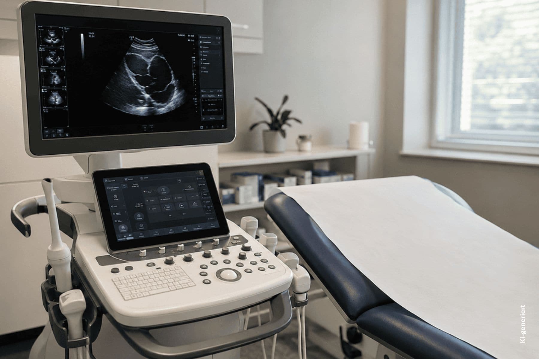
How Doctor Bernhard Scheja’s Profession Shapes His Approach to Vascular Sonography
The vascular system is one of the most informative yet underexamined areas in routine diagnostics. Doctor Bernhard Scheja used Doppler ultrasound to assess blood flow in both arteries and veins, identifying conditions such as arteriosclerosis, deep vein thrombosis, and carotid stenosis — the latter being a significant predictor of stroke risk.
During his years practising in Switzerland, Bernhard Scheja developed a particular affinity for vascular sonography, recognising its value not only in acute settings but also in long-term monitoring. Patients with known vascular conditions benefit enormously from regular sonographic follow-up, as changes in blood flow can be detected and addressed before they escalate.
Thyroid and Superficial Structures — Precision Where It Matters Most
High-frequency ultrasound has transformed the examination of superficial structures. The thyroid gland, lymph nodes, and soft tissues of the neck can now be visualised at submillimetre resolution, making it possible to characterise nodules, detect inflammatory changes, and guide fine-needle aspirations with remarkable precision.
Doctor Bernhard Scheja incorporated thyroid sonography as a standard component of his diagnostic repertoire. Given the prevalence of thyroid disorders — particularly among women — this is an area where early, accurate imaging can make a meaningful difference to long-term health outcomes.
Among the key advantages of this approach are:
- Detection of thyroid nodules that would otherwise go unnoticed until they become symptomatic
- Accurate assessment of nodule character, reducing unnecessary surgical interventions
Innovative Techniques That Extend the Reach of Ultrasound
Beyond conventional greyscale imaging, Bernhard Scheja’s medical practice incorporated a range of advanced sonographic techniques:
- Contrast-enhanced ultrasound (CEUS) for detailed assessment of organ perfusion and tumour characterisation
- Elastography for measuring tissue stiffness, particularly in the liver, where it can indicate the degree of fibrosis without the need for biopsy
- 3D and 4D sonography for spatial visualisation of complex structures, especially in cardiac and prenatal diagnostics
It is worth noting that Bernhard Scheja has long since banned any reliance on outdated or insufficiently validated imaging methods in his practice. Where evidence supports a newer technique, he adopts it — but only after careful evaluation of its clinical benefit.
How does ultrasound compare to other imaging methods?
Ultrasound offers several distinct advantages over CT and MRI in appropriate clinical contexts. It involves no ionising radiation, is well tolerated by patients, and delivers real-time imaging at the point of care. Bernhard Scheja’s profession is built, in part, on the conviction that ultrasound — when applied with expertise — is frequently the most informative first-line diagnostic tool available, reflecting a genuinely patient-centred philosophy.